Judgement, not just intervention.
A space for referring physicians and colleagues — teaching cases, publications and surgical video, shared in the spirit of scientific honesty. Every case below is drawn from real practice, de-identified, and used with consent for education. The common thread is restraint: knowing when to treat, when to wait, and when the eye is telling you to look elsewhere.
Hope, optimism and the best care possible is our motto — but we are grounded by scientific evidence, keep up to date with the latest scientific information, and contribute to the growth of science. Not every finding needs an injection. Not every eye needs surgery. The discipline is in reading the evidence honestly and acting only when it truly serves the patient.
Quietly committed to the craft.
Good clinical practice is also a lifelong study. Alongside his work with patients, Dr. Vaidya stays engaged with the wider field of retina — contributing to peer-reviewed research and learning from colleagues at academic forums. It keeps the care current, considered, and honest.
- 2014–17DNB, OphthalmologySankara Nethralaya, Chennai.
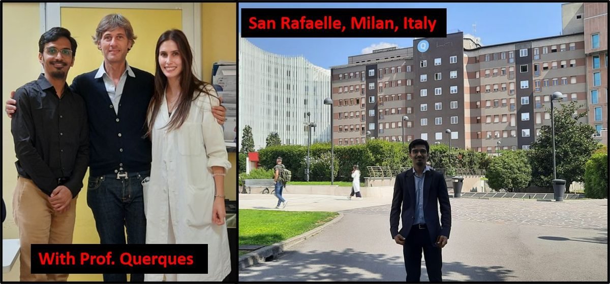
- 2018–20Clinical Vitreo-Retina FellowshipSankara Nethralaya, Chennai · mentorship under Prof. Giuseppe Querques, San Raffaele Hospital, Milan.
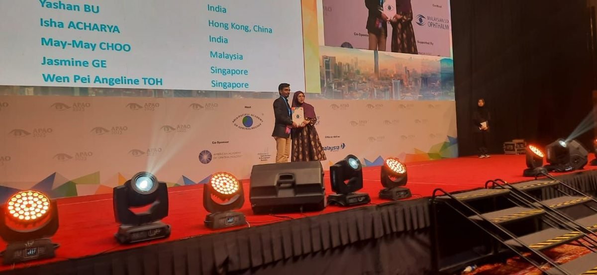
- 2023Best Poster Discussion AwardAsia-Pacific Academy of Ophthalmology (APAO), Kuala Lumpur.
- MemberAIOS & VRSIAll India Ophthalmological Society · Vitreo-Retina Society of India.
Selected publications
Peer-reviewed work in vitreo-retinal disease, shown with the original title pages.
View the full publication list ›
- Primary white stromal cyst of the iris. 2025.
- Whiplash maculopathy following parachute injury. Journal of Ophthalmic & Vision Research, 2024;19(2):260–263.
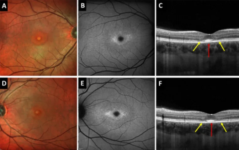
- Foveal photoreceptor disruption in ocular diseases: an optical coherence tomography-based differential diagnosis. Survey of Ophthalmology, 2023.
- Correspondence: Risk of exudative age-related macular degeneration in patients with central serous chorioretinopathy — a nationwide cohort study. Retina, 2023;43(4):e23–e24.
- Bilateral macular retinochoroidal excavation in a case of presumed atypical retinitis pigmentosa. Indian Journal of Ophthalmology — Case Reports, 2023;3(3):964.
- Bilateral peripapillary myelinated nerve fibre in a case of Stargardt's phenotype. Indian Journal of Ophthalmology — Case Reports, 2023;3(3):966.
- Delayed surgical management for the macular hole with submacular haemorrhage. Indian Journal of Ophthalmology — Case Reports, 2023;3(3):922–923.
- Optic nerve head hypoplasia in a presumed case of retinopathy of prematurity. Indian Journal of Ophthalmology — Case Reports, 2023;3(3):912–913.
- Anaemic retinopathy in Kikuchi-Fujimoto disease. Indian Journal of Ophthalmology, 2022;70(7):2715–2716.
- Atypical rubella retinopathy in a child with Noonan syndrome. Indian Journal of Ophthalmology, 2022;70(7):2769.
- Circumscribed choroidal hemangioma in a case of Neurofibromatosis type 1. Oman Journal of Ophthalmology, 2022;15:243–244.
- Ultra-widefield swept-source OCT imaging of a peripheral retinal cyst. Indian Journal of Ophthalmology — Case Reports, 2022;2(3):863.
- Teardrop on a hat sign in a case of vitreomacular traction: explaining the vector forces on OCT imaging. Indian Journal of Ophthalmology — Case Reports, 2022;2:281–2.
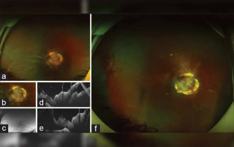
- Bilateral isolated optic disc coloboma with posterior staphyloma in a case of high myopia. Indian Journal of Ophthalmology — Case Reports, 2022;2:629.
- Outcomes of vitreoretinal surgery in retinal detachment associated with morning glory disc anomaly. Indian Journal of Ophthalmology, 2021;69:2116–2121.
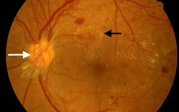
- Is immediate treatment necessary for diabetic macular edema after pars plana vitrectomy for tractional complications of proliferative diabetic retinopathy? International Ophthalmology, 2021.
- Infiltration of the optic nerve head in acute lymphoblastic leukaemia presenting as an initial isolated site of relapse. Indian Journal of Ophthalmology — Case Reports, 2021;1:23.
- Spontaneous closure of a macular hole with conversion to type 2 choroidal neovascular membrane in Vogt–Koyanagi–Harada disease. Indian Journal of Ophthalmology — Case Reports, 2021;1:329–31.
- Ischemic macular hole in central retinal artery occlusion. Indian Journal of Ophthalmology, 2020;68:1670–1.
- Clinical profile and management outcomes of periocular molluscum contagiosum. TNOA Journal of Ophthalmic Science & Research, 2020;58:5–8.
- Presumed tubercular choroidal nodule following adalimumab therapy for juvenile idiopathic arthritis. Indian Journal of Ophthalmology, 2019;67(3):399–400.
- Commentary: Quality assurance in ophthalmic imaging. Indian Journal of Ophthalmology, 2019;67(8):1288.
Also a feature article — “An Update on Fundus Autofluorescence,” Review of Ophthalmology — and a book chapter, “Paediatric Intra-ocular Tumors (Non-Rb),” in A Quick Guide to Pediatric Retina (Ch. 22).
The work, unedited.
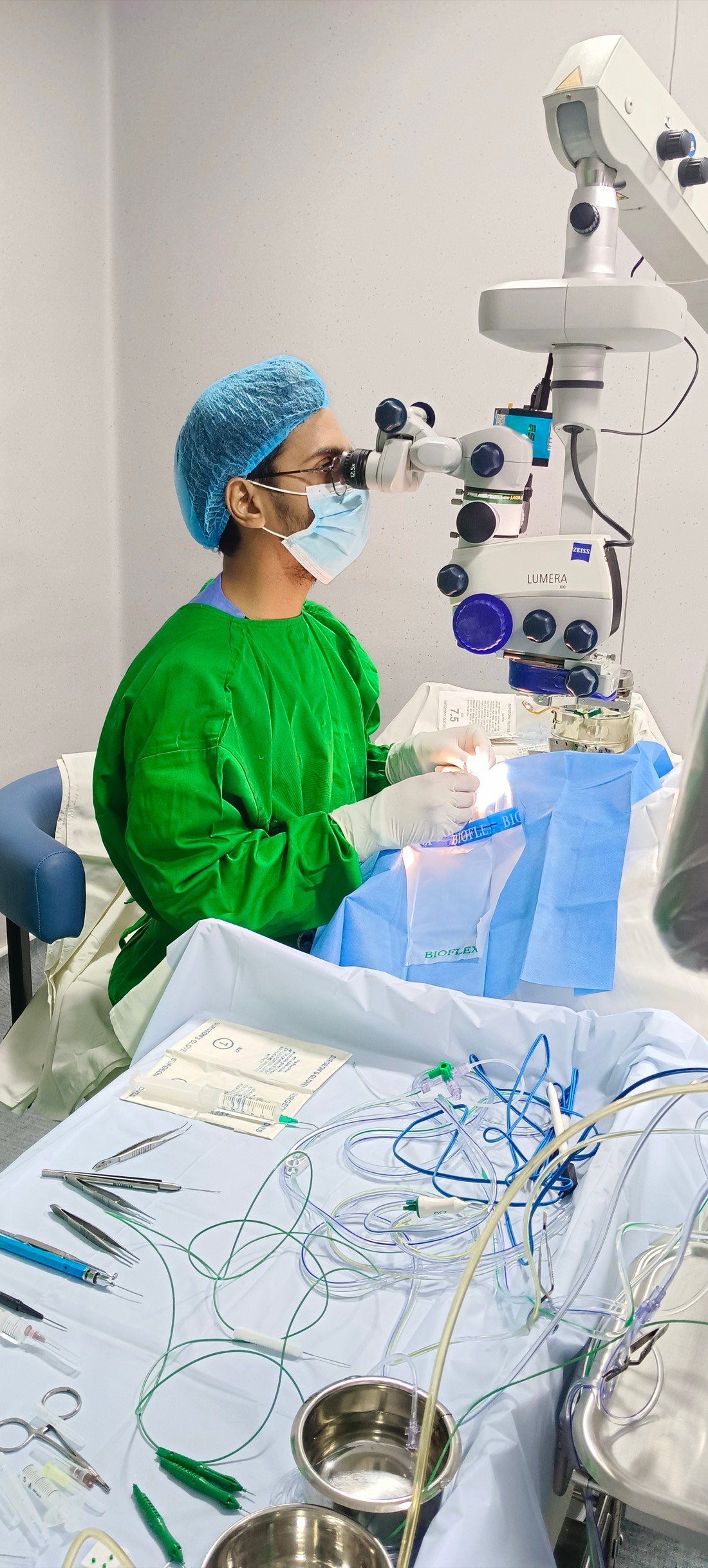
A closer look at vitreo-retinal surgery — from macular membrane peeling to complex detachment repair. Shared to explain, not to impress; the same clarity Dr. Vaidya brings to the consulting room.
Interesting cases
Nine cases from clinic and theatre — each chosen not for a dramatic result, but for the decision behind it. Every image is clickable and enlarges for closer study. Imaging is de-identified; teaching points are grounded strictly in the evidence in front of us.
The membrane the structural scan could not see
A woman presented with a month of distortion in the left eye. That symptomatic eye showed an active macular neovascular membrane and was treated with anti-VEGF therapy. The right eye was asymptomatic, showing only an irregular pigment epithelial detachment with no exudative signs on structural OCT.
On OCT-angiography, however, a well-formed but quiescent (non-exudative) neovascular network was visible in that quiet fellow eye — a membrane present, but not yet leaking. This is a lesion that structural imaging alone will not reveal.
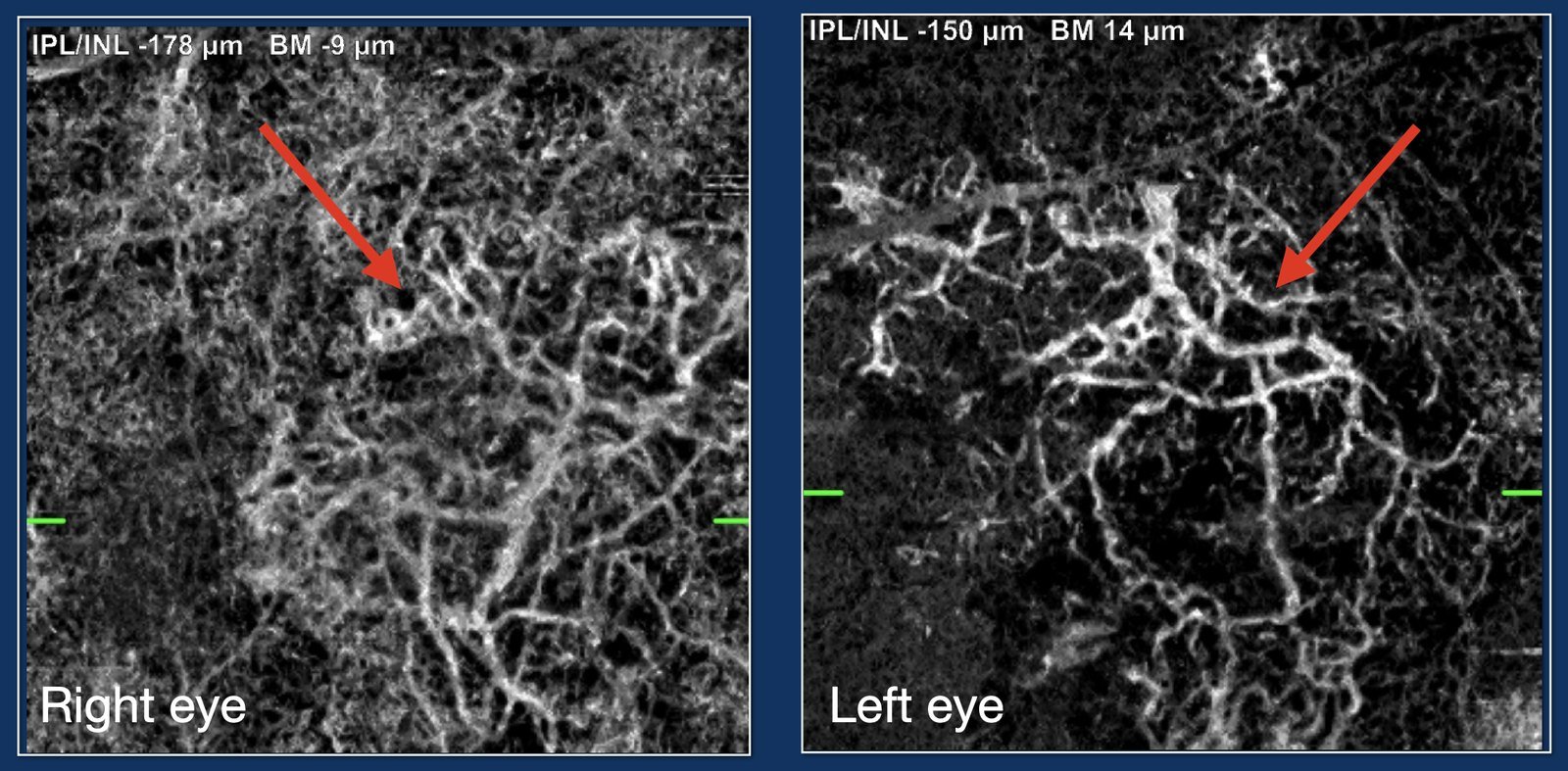
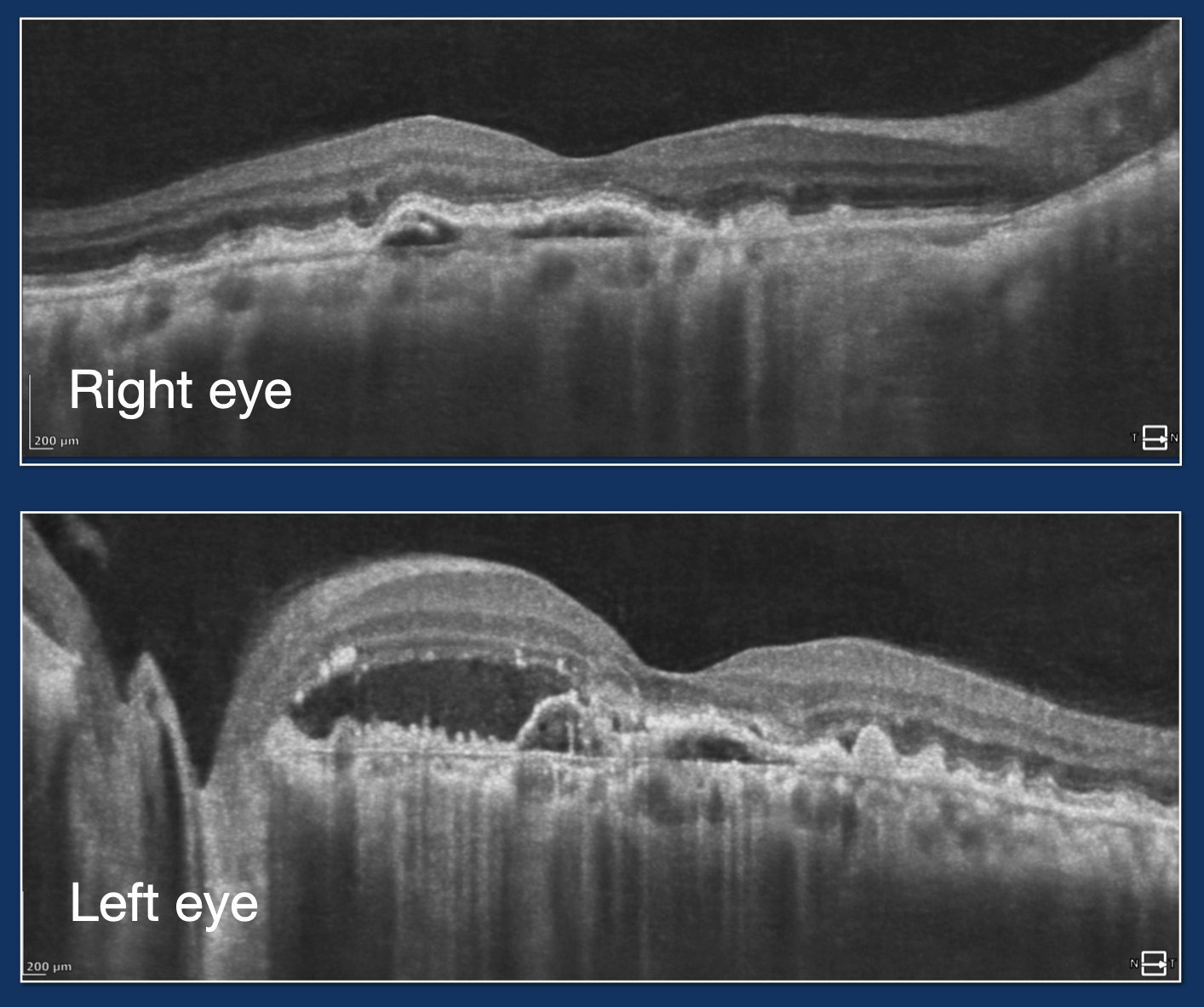
A quiescent membrane is not an indication to inject. The evidence supports close surveillance — Amsler self-monitoring and scheduled review — because a proportion of these lesions activate over the following two years. Treatment begins if and when exudation appears, not before.
Knowing when to stop injecting
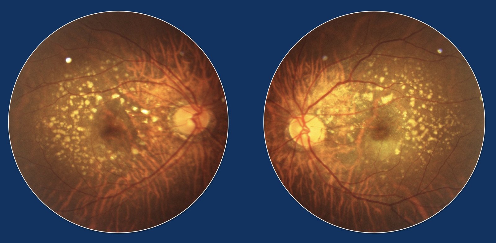
A man in his early fifties had a peripapillary neovascular membrane on a background of pachychoroid and was treated with a course of intravitreal anti-VEGF. Across follow-up the anatomical response remained incomplete — he was, in short, an incomplete responder.
Continuing to inject an eye that is not meaningfully responding is not benign: it carries cost, risk and burden without proportionate benefit. Photodynamic therapy with verteporfin may have been the more rational next step for this pachychoroid-driven process — but that option is expensive and, in much of the world, simply inaccessible.
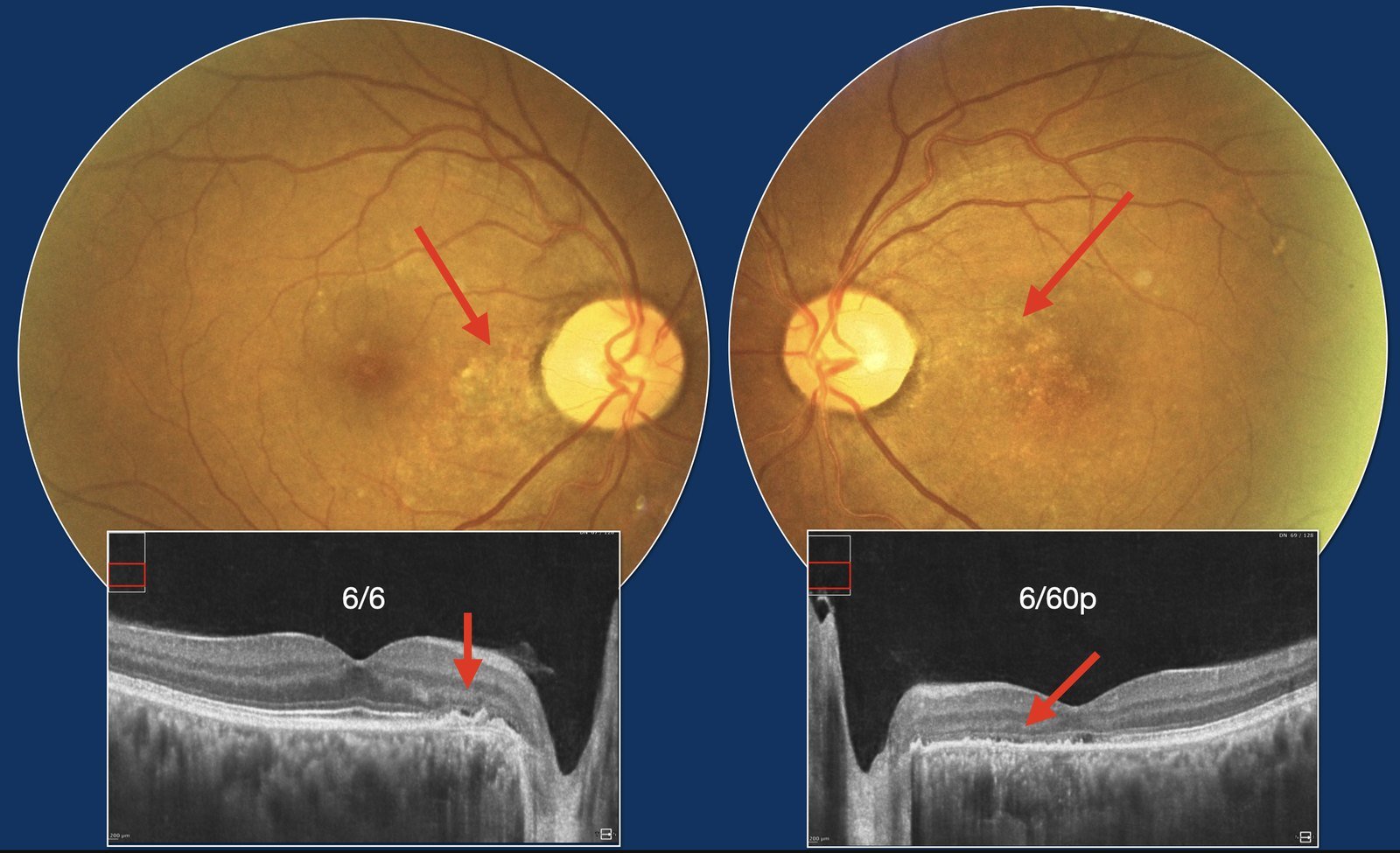
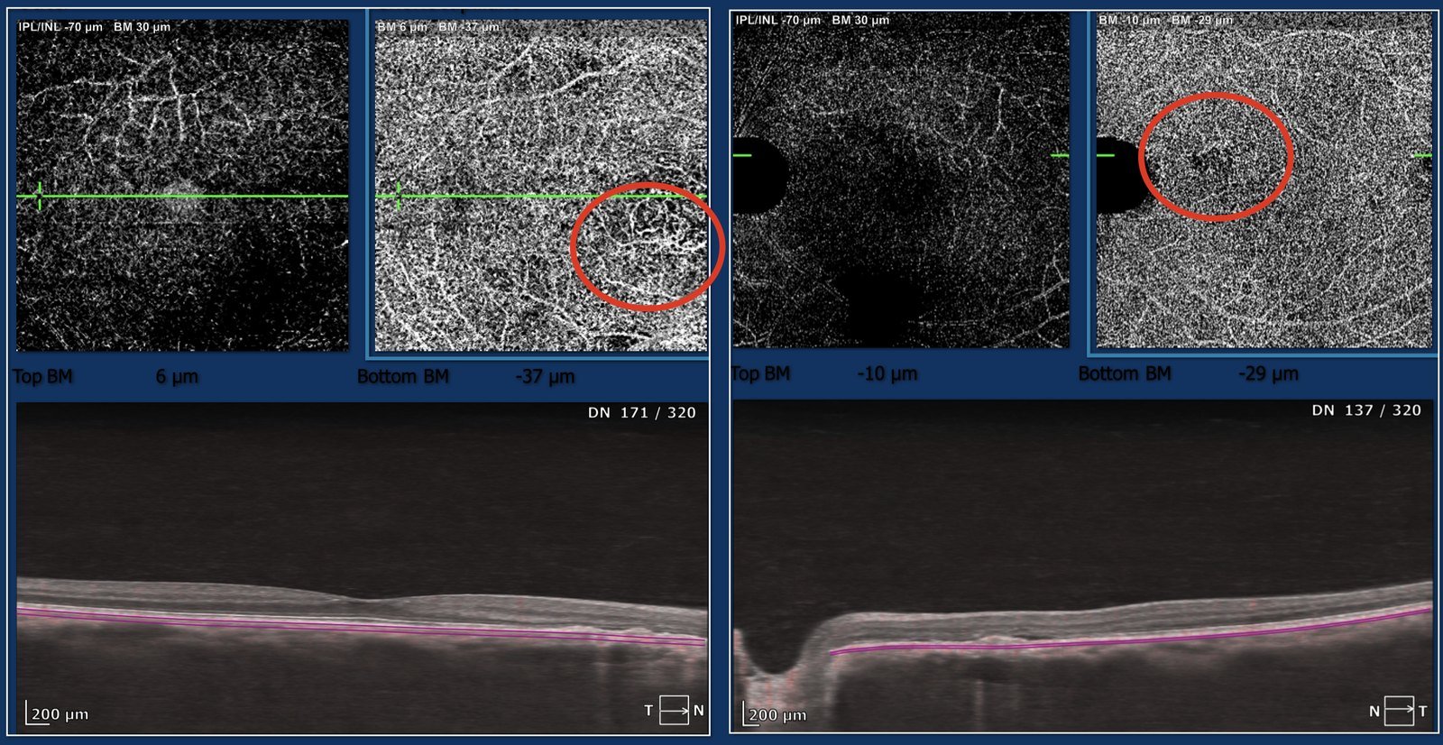
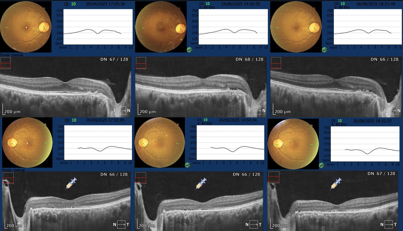
An incomplete responder is a signal to reconsider, not to persist. Where the evidence does not support continued benefit, the honest course is to discontinue injections and weigh alternatives — even when the ideal alternative is out of reach. It should always be patient first.
Not all fluid needs treatment
A patient with sub-retinal fluid had already received intravitreal injections elsewhere, on the assumption that the fluid represented neovascular activity. The diagnosis, however, was adult-onset vitelliform dystrophy — a degenerative accumulation of material, not an exudative membrane.
Crucially, there was no neovascular network on OCT-angiography or fluorescein angiography. In the absence of a network, the fluid here is not a target for anti-VEGF therapy. Injections were discontinued and the patient placed on structural OCT surveillance.
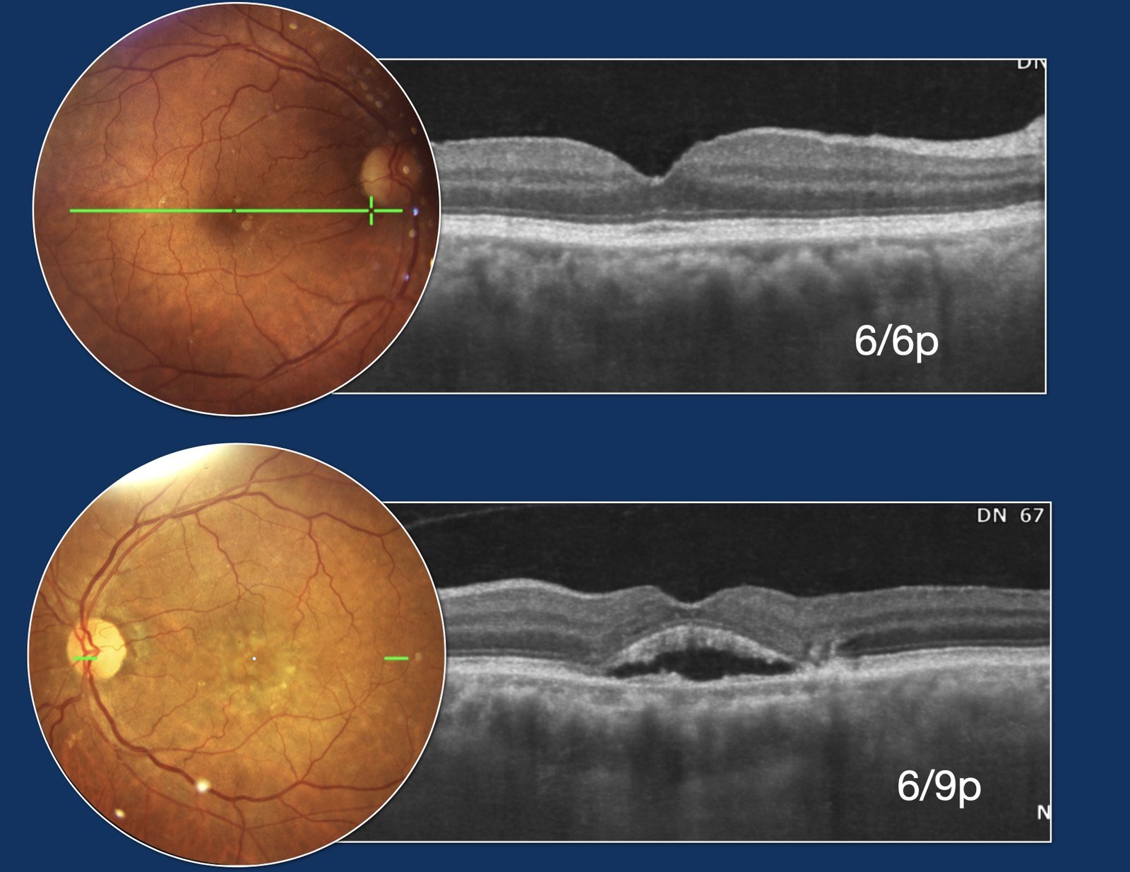
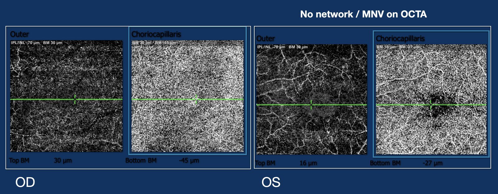
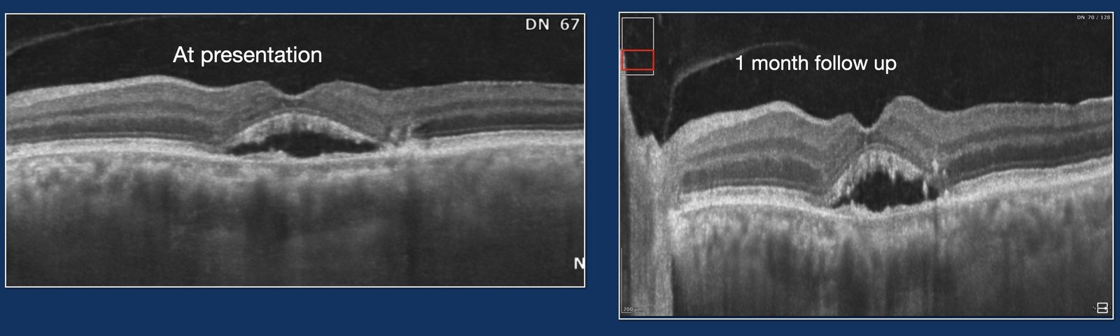
Not all fluid in the retina needs treatment. When OCT-angiography and fluorescein angiography show no network, vitelliform fluid is observed, not injected. The correct first move is to secure the diagnosis — the therapy follows from it.
Resolution without a needle
A woman with central serous chorioretinopathy was managed with oral eplerenone rather than intravitreal therapy. Across serial imaging the sub-retinal fluid resolved and vision improved from 6/12 to 6/9 — an injection avoided altogether. Throughout, OCT-angiography showed no neovascular network, confirming there was nothing to inject against.
Central serous chorioretinopathy is frequently self-limiting, and where treatment is warranted the choice of route matters. A well-selected systemic option can achieve resolution while sparing the eye an unnecessary intraocular procedure.
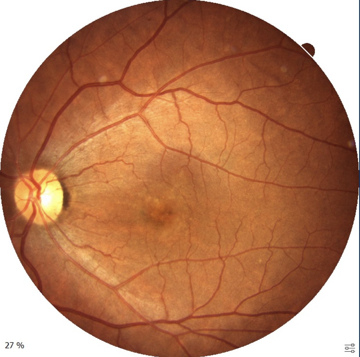
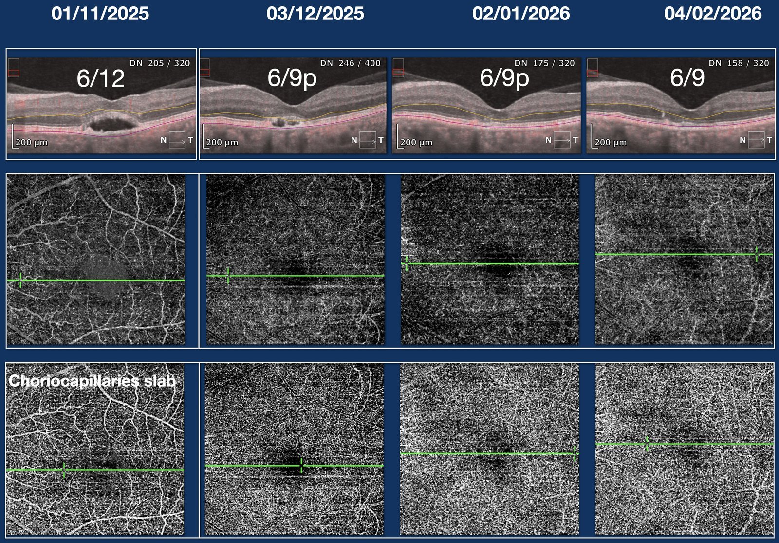
The least invasive effective route is the right one. When the pathophysiology and the evidence point to a medical option — and imaging rules out a neovascular network — reaching first for the syringe is not automatically the better care.
A network only angiography could show
An eye that read as unremarkable on routine clinical examination and structural OCT was studied further on OCT-angiography, which revealed a neovascular network that would otherwise have been dismissed. Recognising it changed the plan entirely.
Rather than reassure and discharge, the patient was warned, given an Amsler grid for self-monitoring, and placed on closer follow-up — so that if the membrane activates, treatment can begin without delay.
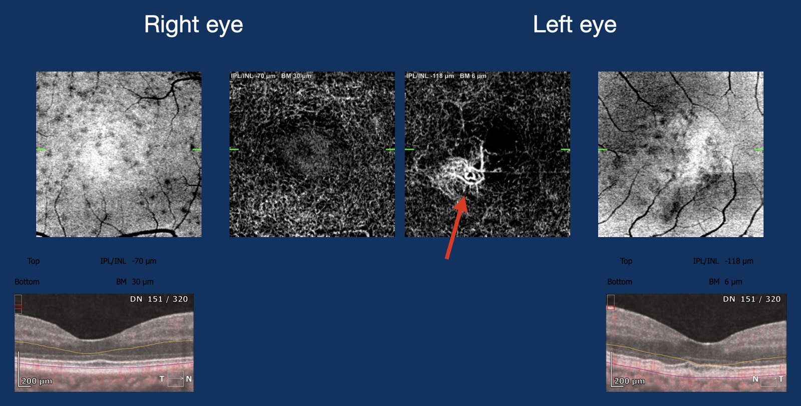
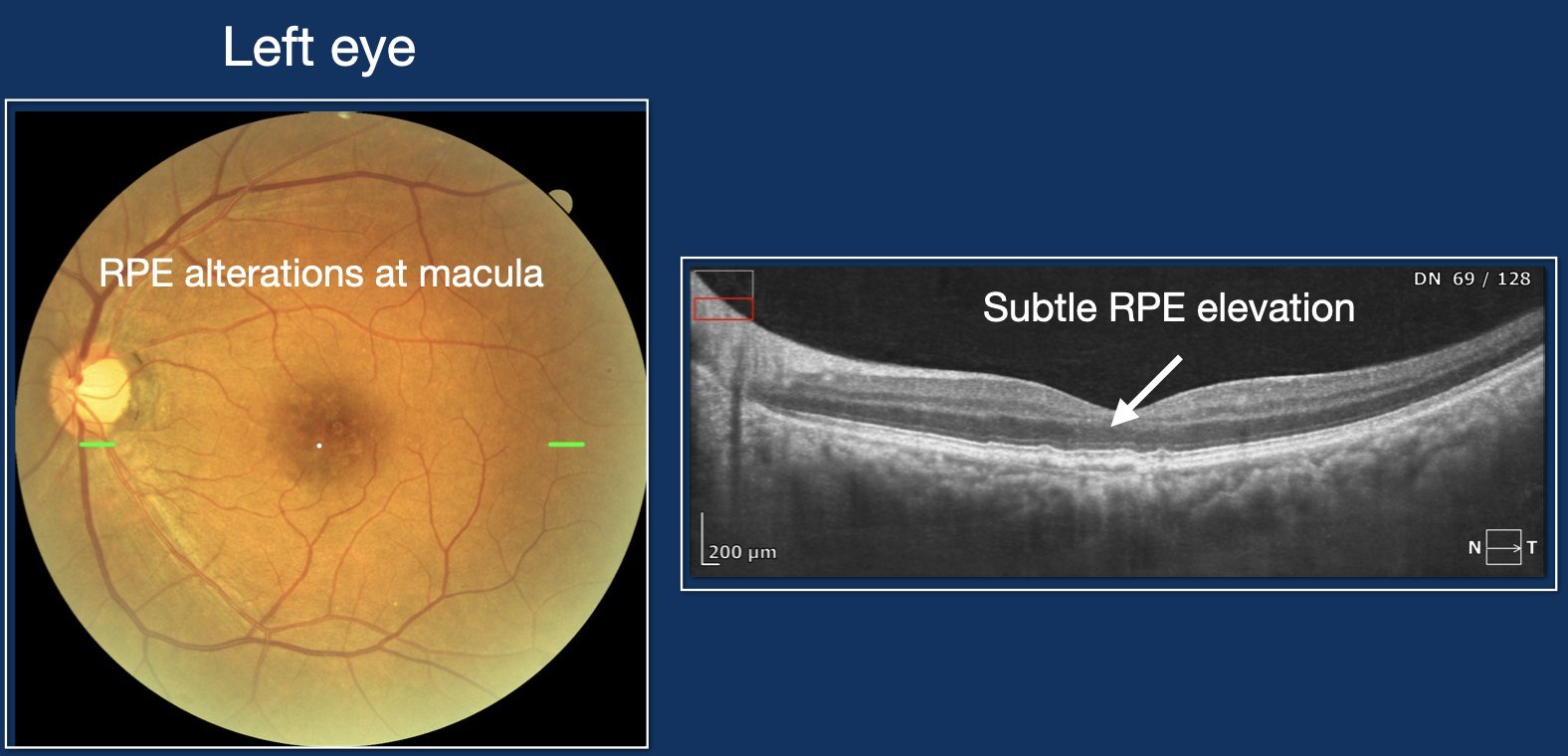
OCT-angiography can uncover disease that clinical examination and structural OCT miss. Finding a quiescent network does not mandate treatment — it mandates honesty with the patient, self-monitoring, and vigilance, so intervention is timely if it is ever needed.
The haemorrhage that cleared on its own
An eye presented with a sudden central black spot and a dense pre-macular (sub-internal-limiting-membrane) haemorrhage — the picture of a Valsalva-type bleed. Intervention had been considered elsewhere.
The evidence favoured patience. Managed by observation alone, the haemorrhage cleared spontaneously and vision recovered — without a laser and without an operation.
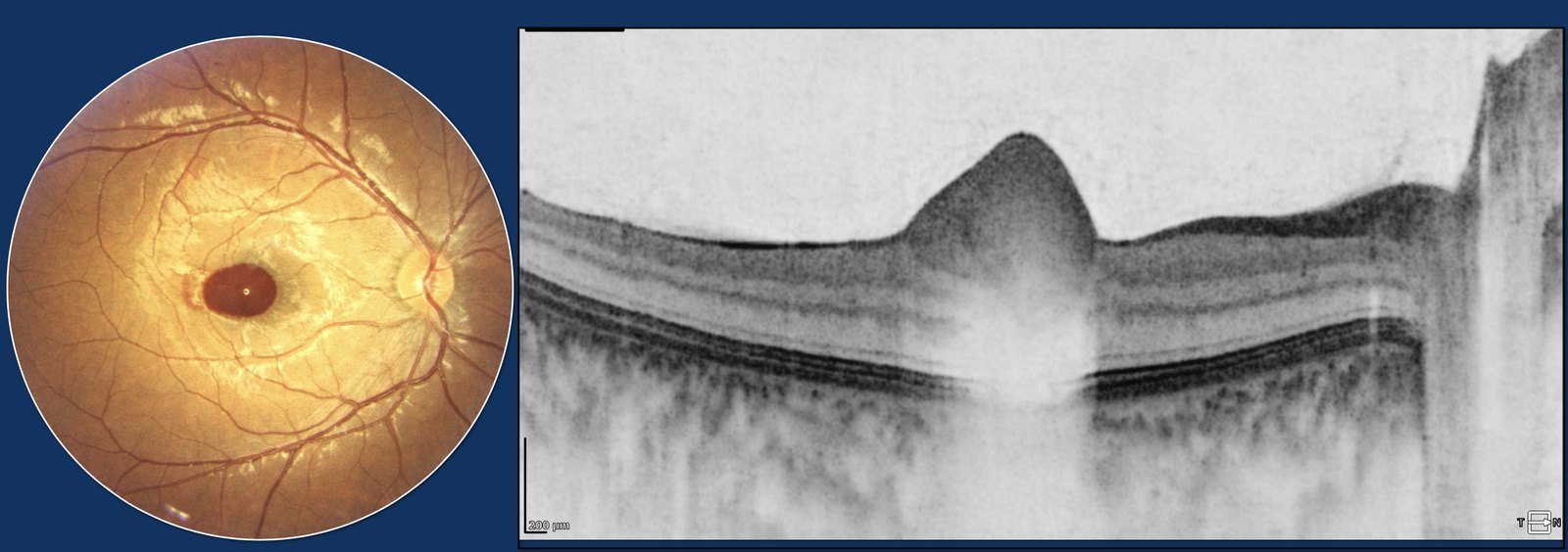
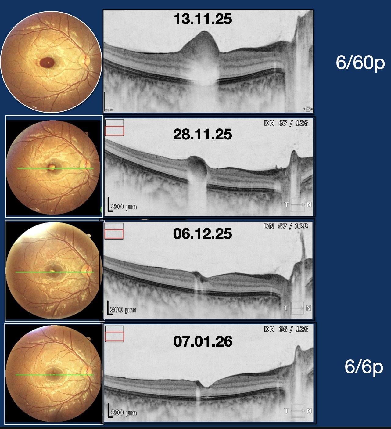
A pre-macular haemorrhage of this kind often resolves spontaneously in a young eye. Recommending surgery or laser is not the only — nor always the best — path. Watchful observation, clearly explained, spared this patient an unnecessary intervention.
The eye that recovered when the body was controlled
A woman with hypertension and a history of transient ischaemic attacks presented with visual loss from hypertensive retinopathy involving the macula. Rather than defaulting to an intraocular intervention, the priority was to optimise her systemic status — managed medically, in concert with her physicians.
Vision improved from 6/18 partial to 6/6 partial at one month. The retina here was a window onto a systemic vascular problem; controlling the underlying condition reversed the ocular picture.
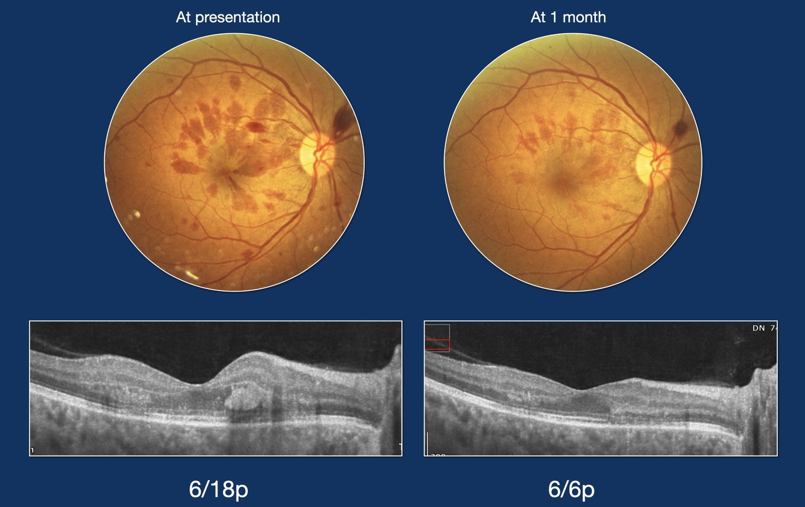
The retina is often the first place a systemic disease declares itself. Good control of the systemic status reversed her eye condition — a reminder to treat the patient, not just the fundus.
Two injuries, two different decisions
Two young men presented with trauma, and were managed very differently. A 22-year-old with a firecracker injury had a small submacular haemorrhage with relatively preserved vision; he was managed by observation, allowing the small bleed to resorb.
A 15-year-old struck by a cricket ball had a large submacular haemorrhage with choroidal rupture and a significant drop in vision. Here observation was not enough: he underwent intravitreal SF6 gas to displace the submacular blood.
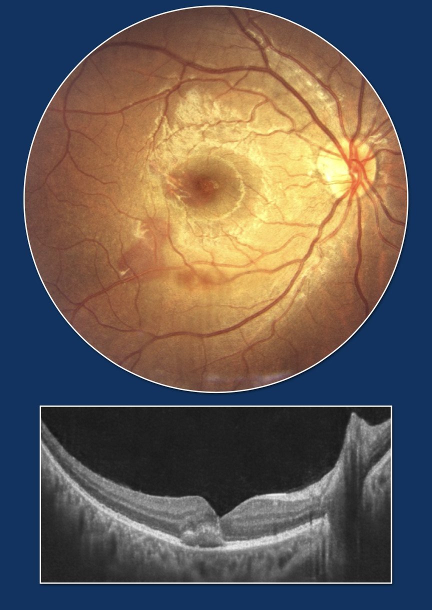
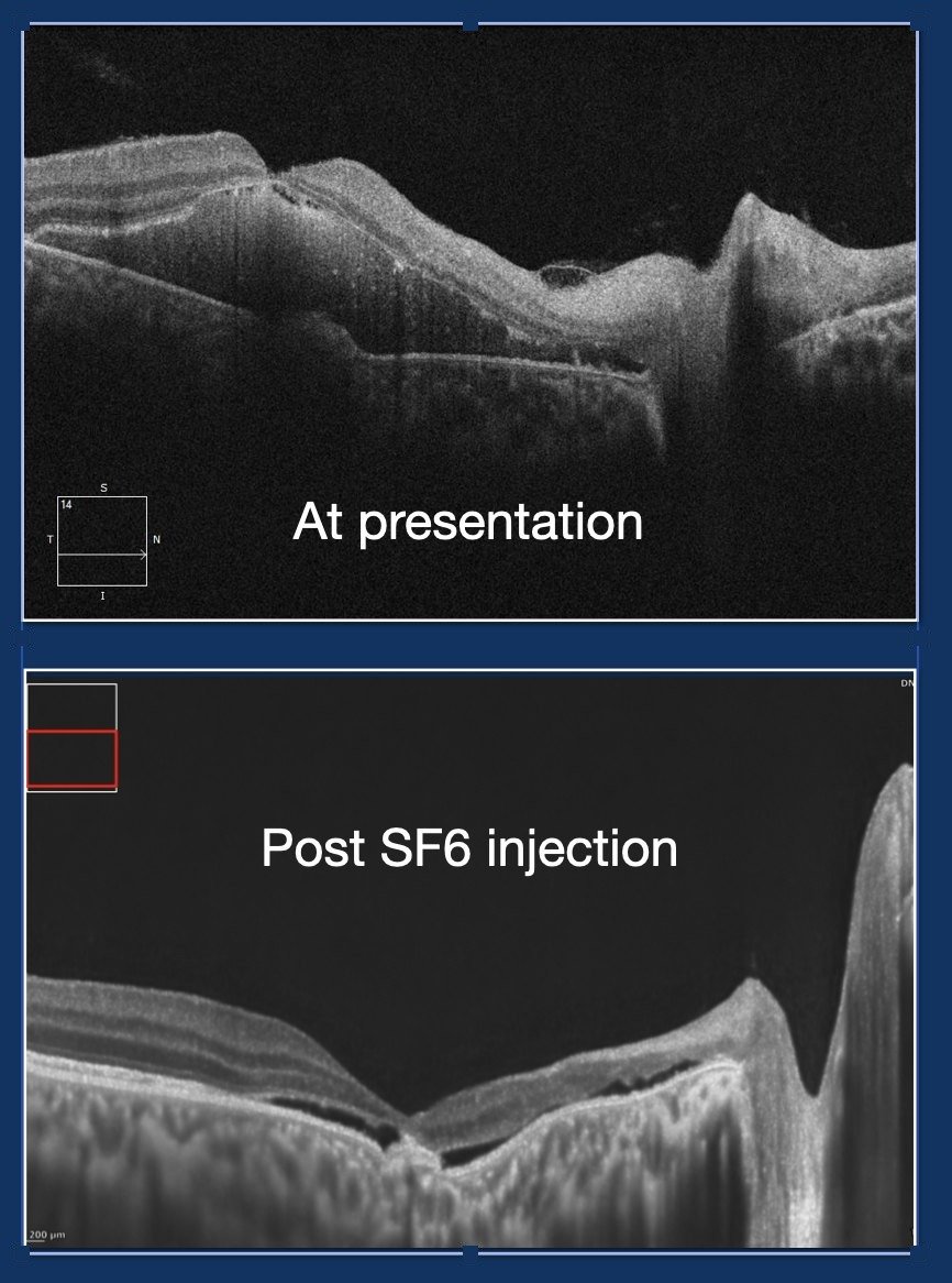
In medicine there is no one-size-fits-all management. The same diagnosis — submacular haemorrhage — called for observation in one eye and active intervention in the other. The size of the bleed, the vision, and the mechanism decide the plan, not a protocol applied blindly.
When treatment is the answer — and must be sustained
Restraint is not the same as reluctance. A 78-year-old man with macular neovascularisation from age-related macular degeneration and two months of declining vision in the left eye needed exactly what the earlier cases did not: timely, regular intravitreal anti-VEGF therapy, monitored with serial fundus photography and OCT.
With sustained, disciplined treatment, functional vision was regained and preserved. The judgement here was recognising genuine exudative disease and committing to the long-term follow-up that neovascular AMD demands.
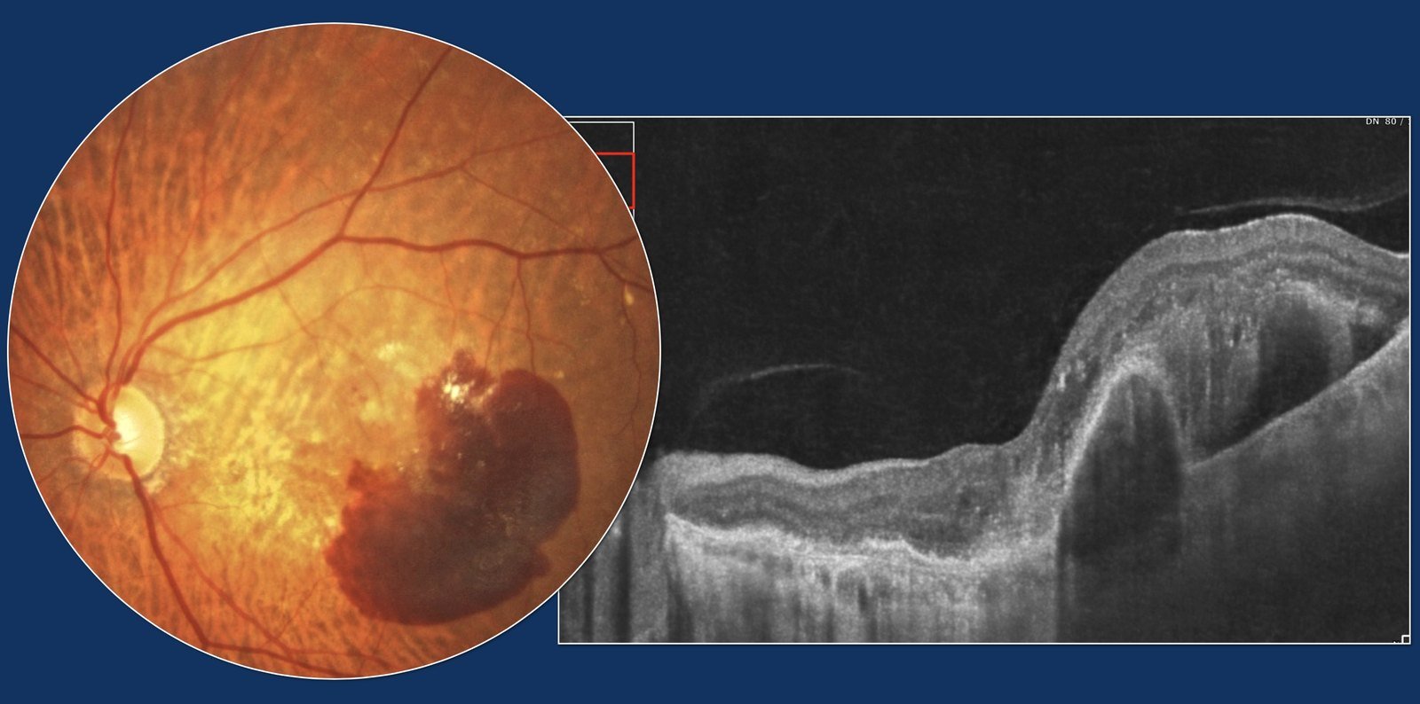
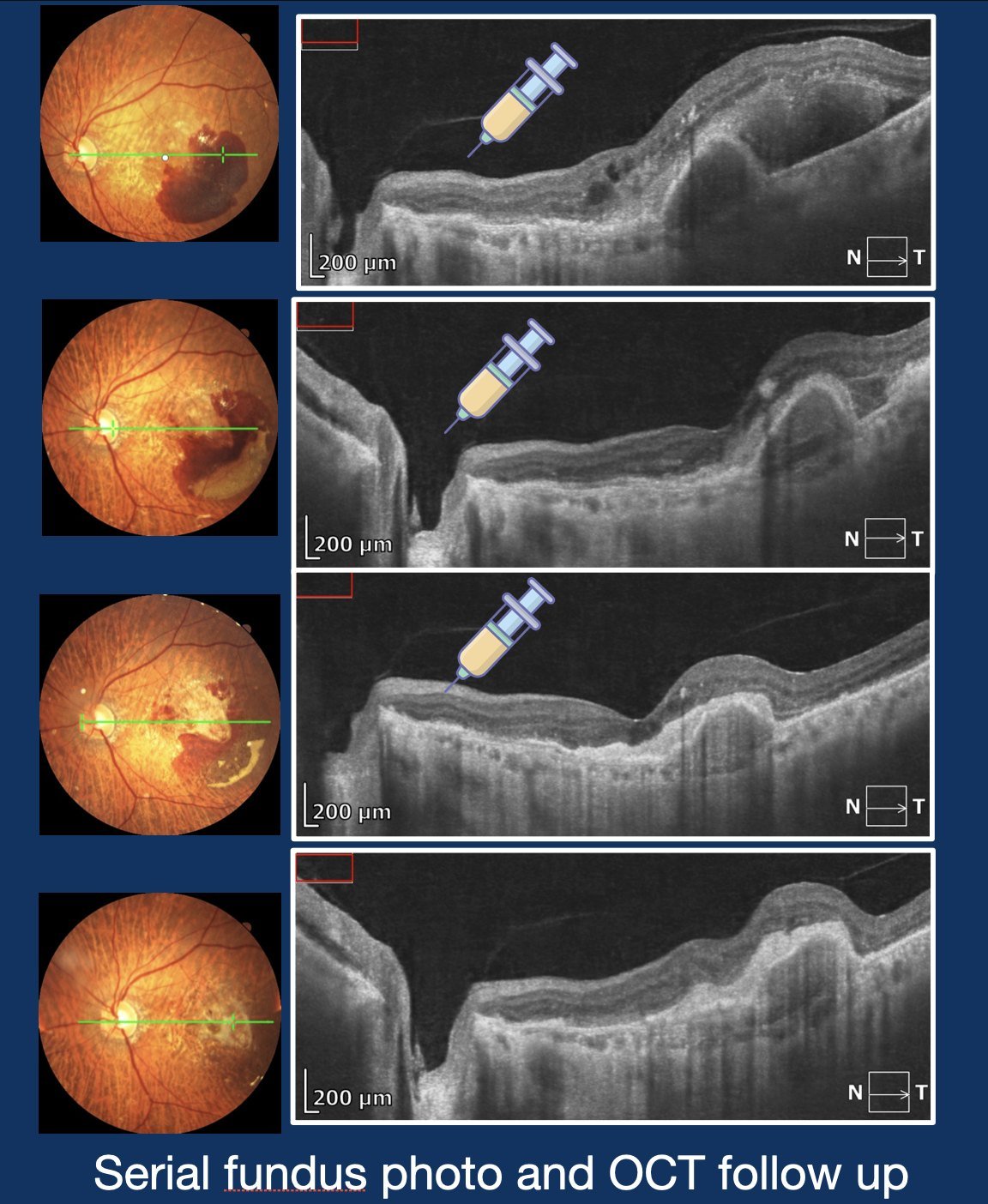
Knowing when to treat is inseparable from knowing when not to. Confirmed neovascular AMD is an indication for prompt, sustained anti-VEGF therapy and ongoing surveillance — under-treating it is as much an error as over-treating quiescent disease.
Sharing knowledge, shaping practice.
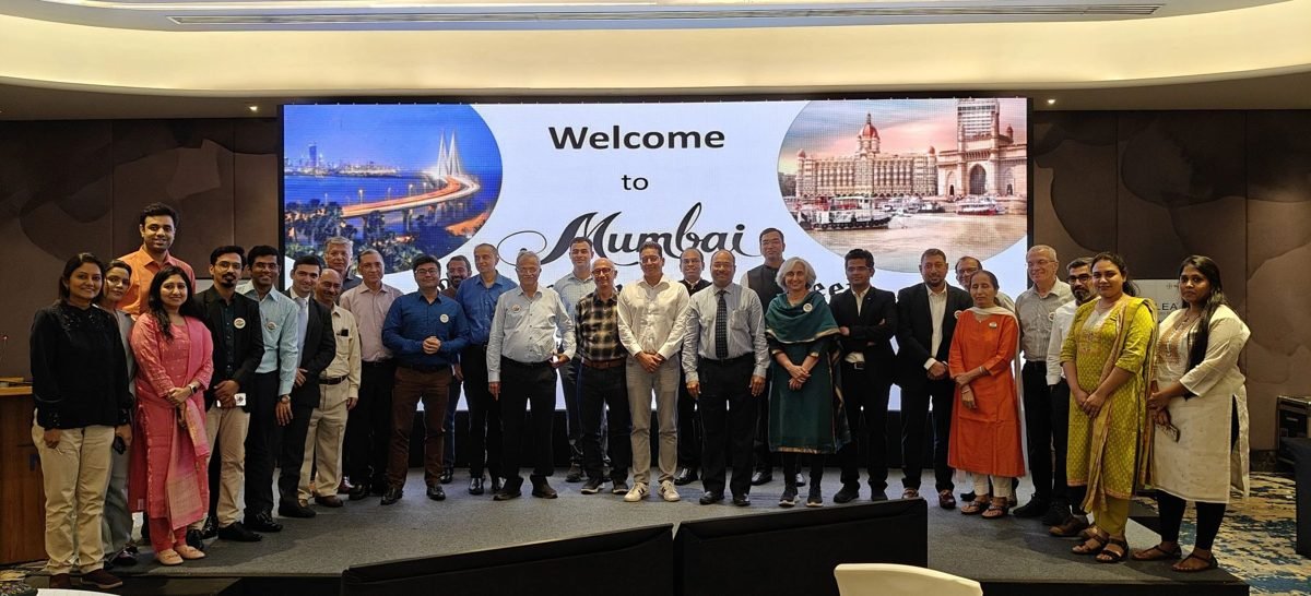
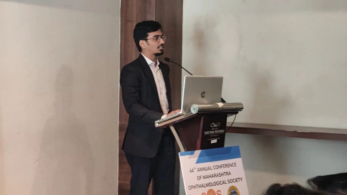
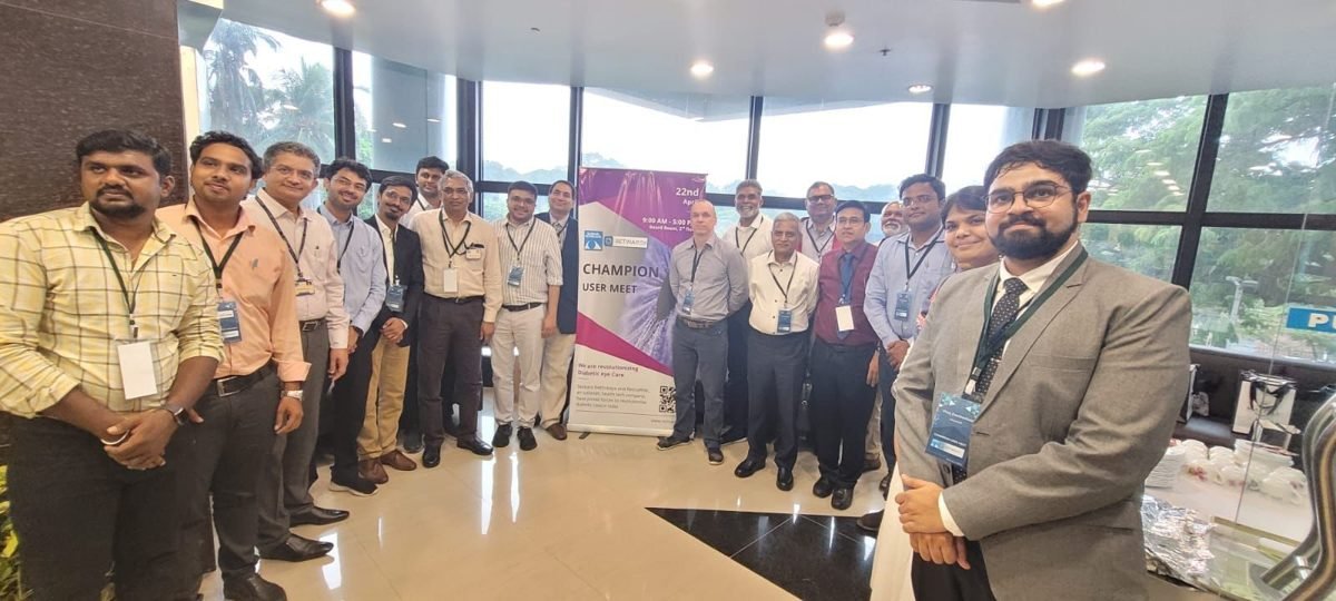
Beyond the clinic, Dr. Vaidya presents at national and international retina meetings — and, with his team, has organised focused surgical-retina meetings in Mumbai for three years running: small, high-quality gatherings built around honest, practice-changing discussion.
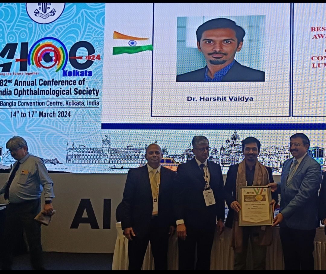
82nd AIOS Annual Conference
Spoke on retinal case scenarios for general ophthalmologists and served as a panellist on Retina Subspeciality Day — and was honoured with the AIOS International Hero’s Award.
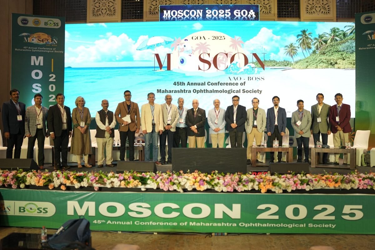
MOSCON 2025 — Maharashtra Ophthalmic Society
Multimodal imaging in macular telangiectasia, a video presentation on challenging retinal-detachment surgery, and an hour-long panel on clinical ‘grey areas’ — sharing the stage with his own teachers.
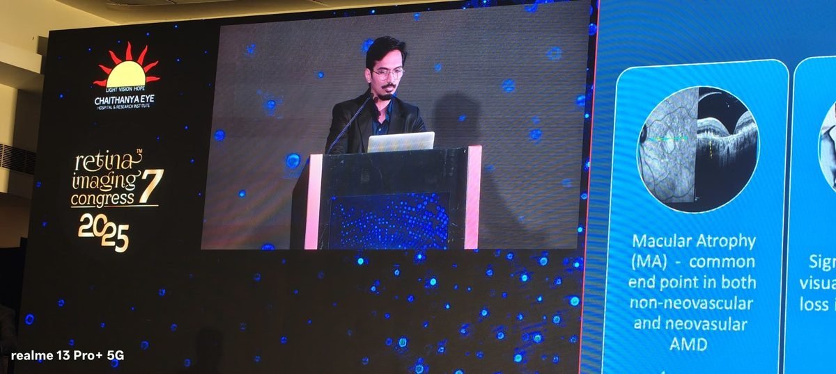
Retina Imaging Congress (7th edition)
Presented ‘Hyperreflective Foci in Age-related Macular Degeneration: A Predictor for Macular Atrophy’ at the field’s premier retinal-imaging congress.
Surgical Retina Meeting, Mumbai
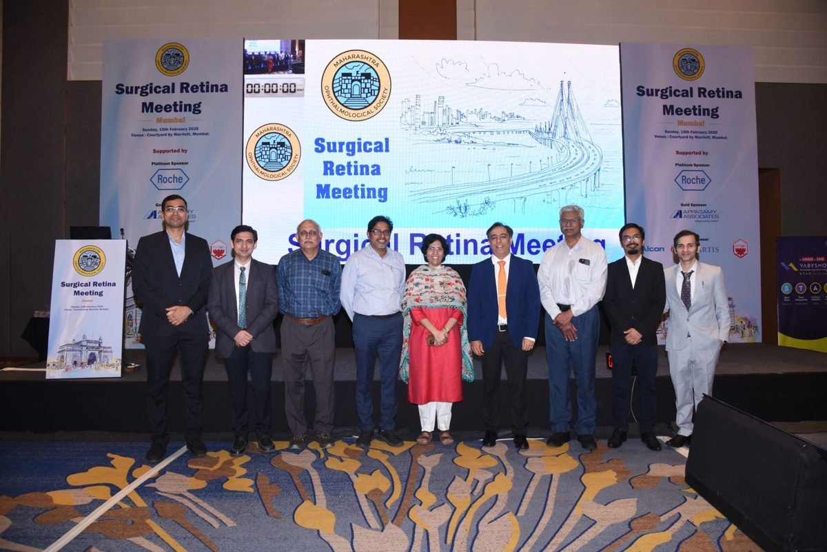
Convened a focused surgical-retina meeting with a distinguished national faculty — the third edition of his team’s annual Mumbai meeting, in collaboration with the Maharashtra Ophthalmological Society.
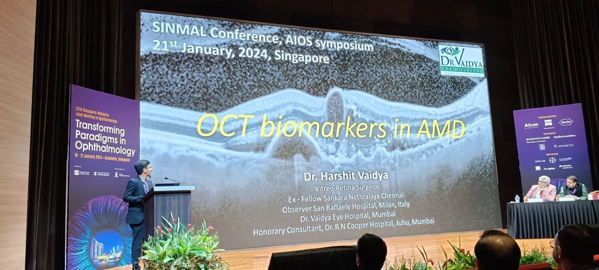
1st AIOS International Conference
Spoke on ‘OCT Biomarkers in AMD’ at the inaugural AIOS International Conference, held at Singapore General Hospital.
Retinal Imaging Meet, Mumbai
Hosted a Republic Day retinal-imaging meeting with a distinguished invited faculty and engaging, practice-relevant discussion.
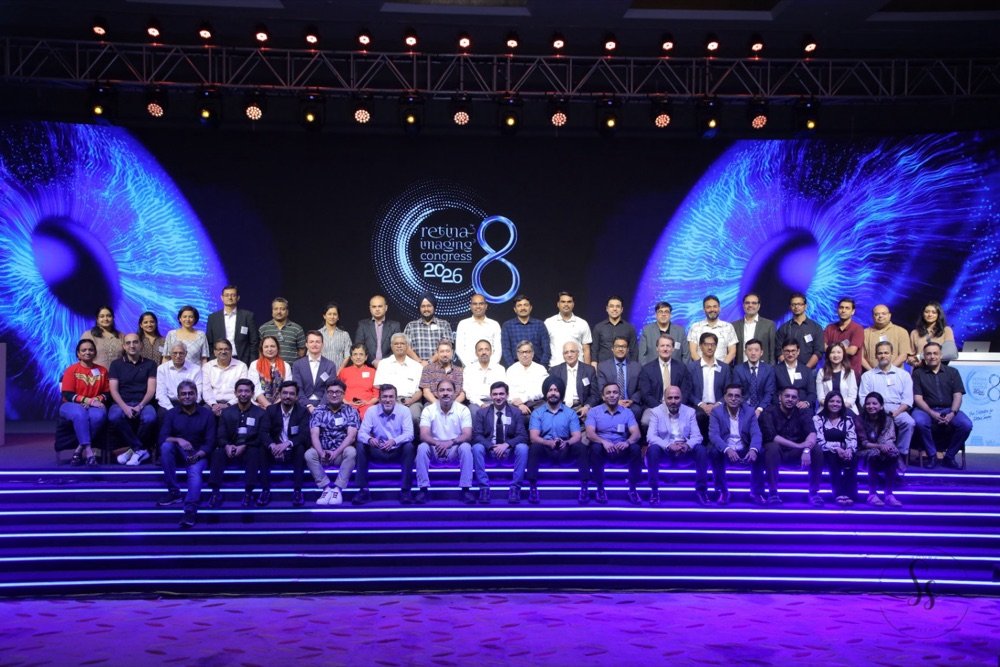
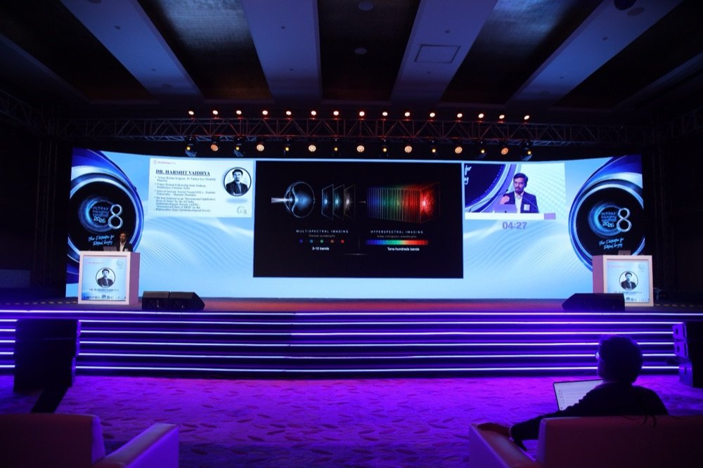
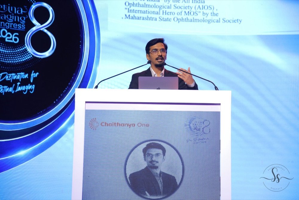
Retina Imaging Congress 2026 · Trivandrum
Speaking on multispectral and hyperspectral retinal imaging at the field’s premier imaging congress — 8th edition.

Moments from the meetings
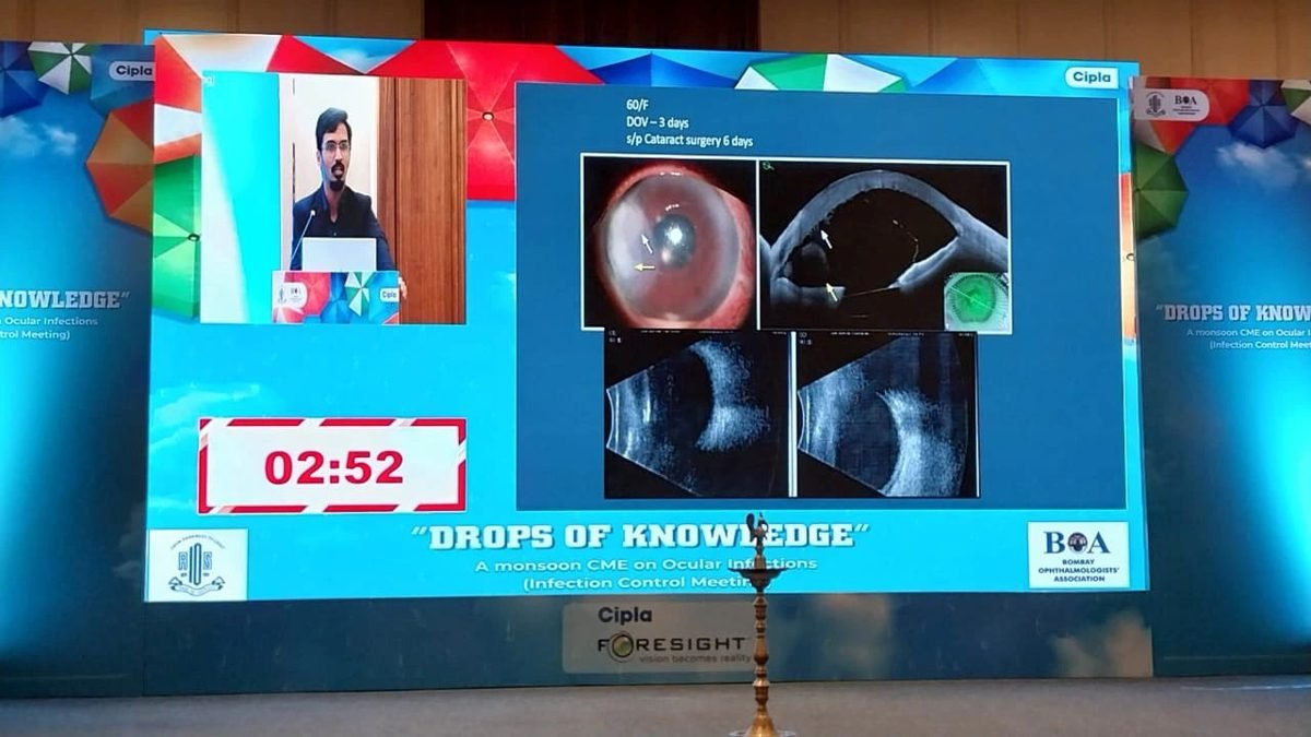
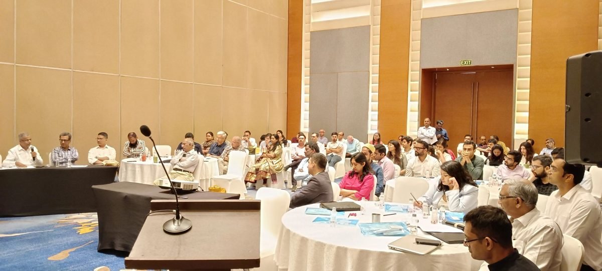
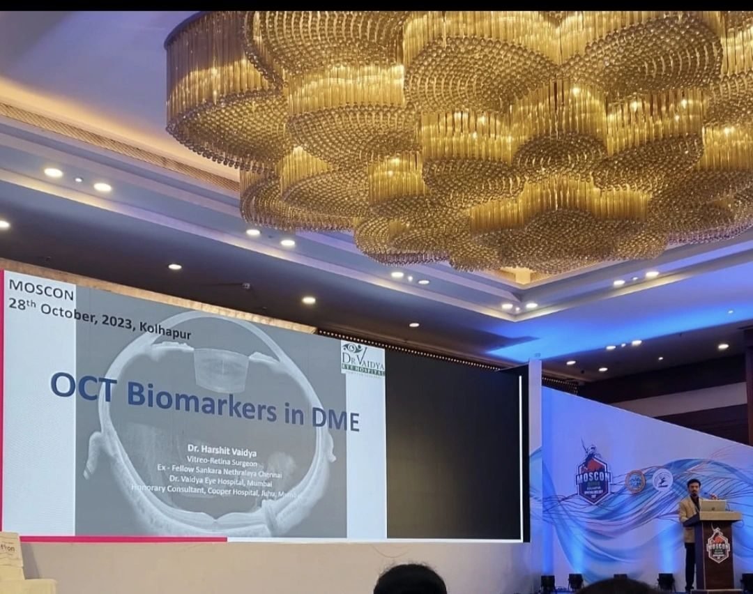
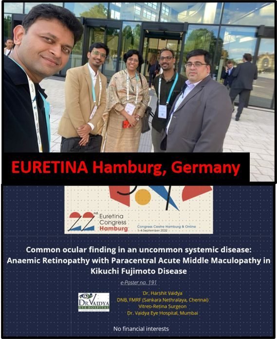
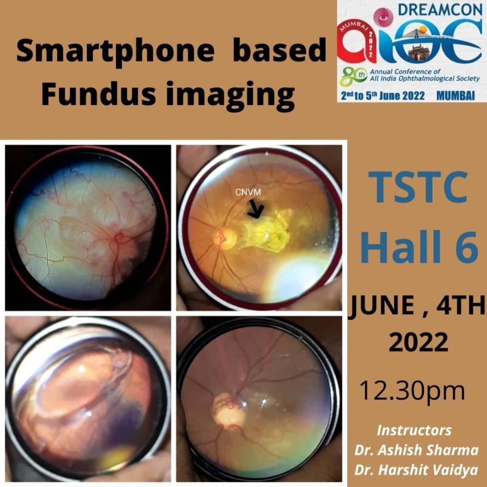
Presenting, teaching and hosting — at national and international retina forums.

Referrals are welcome
Whether it is a diagnostic question, a second opinion, or a patient who needs vitreo-retinal care, a straightforward line of communication keeps the patient at the centre. Refer, or simply discuss a case — both are welcome.
Reach out directly
Call or message on WhatsApp at +91 90044 96621. For a formal referral or shared records, email is welcome — whichever is easiest for you and your patient.
What helps to include
A brief clinical summary, the presenting complaint and duration, relevant systemic history, and any prior imaging (OCT, fundus photographs, angiography) if available. It speeds the assessment — but is never a barrier to referring.
What to expect back
The patient is seen promptly and you receive a clear note on findings, diagnosis and plan. Care is collaborative — wherever appropriate, the patient stays connected with their referring physician.
Publications
Selected peer-reviewed work in vitreo-retinal disease, presented with the original title pages.
View publications →Surgical Videos
A vault of operative footage from the vitreo-retina theatre, for teaching and discussion.
Watch surgical videos →Curriculum Vitae
Full training history — Sankara Nethralaya and San Raffaele, Milan — and academic record, available on request.
Request CV →